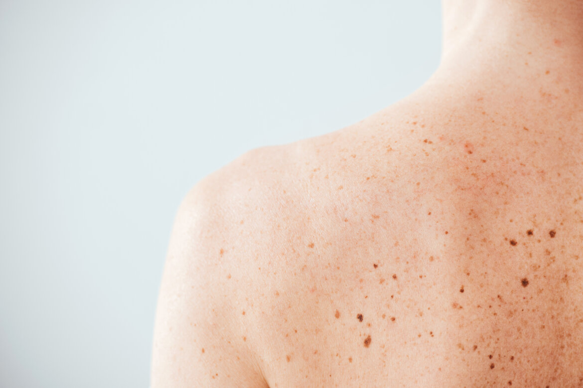
CUTANEOUS AND SUBCUTANEOUS CANCERS

Cutaneous and Subcutaneous Tumours: Overview, Diagnosis, and Treatment
Cutaneous and subcutaneous tumours are medical conditions in which the cells of the skin and subcutaneous tissues grow abnormally. These tumours can be benign or malignant and may occur in different parts of the body.
Types of Cutaneous and Subcutaneous Tumours
Cutaneous tumours include several types, such as basal cell carcinomas (BCCs, the most common skin cancer), squamous cell carcinomas (SCCs, the second most common), and melanomas (the most dangerous skin cancer). On the other hand, subcutaneous tumours can include lipomas, fibromas, and neurofibromas.
Causes and Prevention
Cutaneous and subcutaneous tumours are often caused by a combination of environmental and genetic factors, such as sun exposure, age, sex, family history, and immunodeficiency. Some of these tumours can be prevented with proper sun protection and early diagnosis.
Diagnosis and Treatment
The diagnosis of cutaneous and subcutaneous tumours is made through a biopsy, in which a tissue sample is taken for microscopic analysis. Treatment depends on the type, location, and severity of the tumour and may involve surgical excision, radiotherapy, or chemotherapy.
Surgical intervention is one of the most effective methods for removing cutaneous tumours. During the procedure, the surgeon removes the tumour along with healthy surrounding tissue to ensure complete elimination. The type of surgical procedure depends on the size and location of the tumour. In plastic surgery, patients can benefit from advanced techniques for the removal of cutaneous tumours.
Surgery can be performed using methods such as standard excision or excision followed by reconstruction with flaps and/or grafts to restore the functionality and aesthetics of the treated area. The choice of treatment depends on the characteristics and location of the tumour.
Recovery and Follow-Up
Postoperative recovery is usually brief, and patients can return to their daily activities within a few days. However, it is essential to follow the surgeon’s instructions to ensure proper healing and prevent complications.
It is important to note that many cutaneous and subcutaneous tumours are curable if diagnosed and treated early. Therefore, consulting a healthcare professional if suspicious skin lesions or changes in a pre-existing mole's colour, shape, or size appear is highly recommended.
Basal Cell Carcinoma (BCC) – The Most Common Skin Cancer
Basal cell carcinoma (BCC) is the most common form of skin cancer and is the most frequently diagnosed among all cancer types. Over 45,000 new cases are diagnosed each year. BCCs result from the abnormal and uncontrolled growth of basal cells in the epidermis, one of the three primary cell types in the skin's upper layer.
Due to its slow growth, most BCCs are treatable and cause only local damage if diagnosed and treated early. Understanding the causes, risk factors, and warning signs of BCC can aid in early diagnosis. The earlier they are diagnosed, the easier they are to treat and cure.
Clinical Presentation of BCC
BCCs can present as open sores (ulcus rodens), red patches, pink growths, shiny bumps, scars, or growths with slightly elevated and rounded edges and/or a central depression. Sometimes, they may become secondarily infected, form scabs, itch, or bleed. Lesions typically occur in sun-exposed areas of the body. In patients with darker skin, approximately half of BCCs are pigmented (i.e., brown).
It is important to note that BCCs can look quite different from person to person.
Are BCCs Dangerous?
Although BCCs rarely spread beyond the primary tumour site, if left to grow, they can lead to disfiguring and dangerous lesions. Untreated BCCs can become locally invasive, expanding in width and depth, and destroy skin, tissues, and bones. The longer a BCC is left untreated, the higher the likelihood of recurrence, sometimes multiple.
A BCC that spreads to other parts of the body becomes particularly aggressive. In even rarer cases, this type of BCC can become potentially fatal.
Early Diagnosis
Thanks to early diagnosis and treatment, nearly all BCCs can be successfully removed without complications. Regular visits to a dermatologist and self-examination for any new, unusual-looking lesions allow for the early detection of skin cancers such as BCC, making them easier to treat.
BCC Warning Signs
Check for BCCs in sun-exposed skin areas. They often present as:
BCCs Can Be Difficult to Diagnose
Remember that BCCs may also appear differently than described. In some individuals, BCCs can resemble non-cancerous skin conditions such as psoriasis or eczema.
If in doubt, consult your trusted dermatologist.
Follow-Up Care
If you have had BCC, there is a higher likelihood of developing another, particularly in the same sun-damaged area or nearby. A BCC may recur even if carefully removed the first time, as some cancer cells might be undetected during surgery. Therefore, it is crucial to have an annual check-up with your dermatologist.
Squamous Cell Carcinoma (SCC) – The Second Most Common Skin Cancer
Squamous cell carcinoma (SCC) of the skin, or cutaneous squamous cell carcinoma, is the second most common type of skin cancer. It is characterised by abnormal and accelerated growth of squamous cells. When detected early, most SCCs are treatable.
Clinical Presentation of SCC
SCCs can appear as scaly red patches, open sores, warty-like lesions, or raised nodules with a central depression. In some cases, SCCs may form superficial crusts, itch, or bleed. These lesions most commonly occur on sun-exposed areas of the body but may also arise in other regions, including the genitals.
How Dangerous is SCC?
While most SCCs can be easily and successfully treated, if left untreated, these lesions can become disfiguring, dangerous, and even life-threatening. Untreated SCCs are prone to local and systemic invasiveness, growing deeper into the skin and spreading to other parts of the body.
SCC Warning Signs
In more advanced stages, these skin cancers can become dangerous. Cutaneous squamous cell carcinoma can develop anywhere on the body but is most commonly found on areas exposed to UV radiation, such as the face, lips, ears, scalp, shoulders, neck, back of the hands, and forearms. SCCs can develop on scars, skin ulcers, and other areas of damaged skin (Marjolin's Ulcer). The surrounding skin often shows signs of sun damage, such as wrinkles, pigment changes, and loss of elasticity.
SCCs may appear as thickened, rough, scaly patches that can crust or bleed. They may also appear as warty-like growths or sores that never fully heal. In some cases, SCCs present as raised growths with a typical central ulcer or depression that may bleed or itch.
Follow-Up Care
Ensure you have at least one annual check-up with your trusted dermatologist.
Melanoma – The Most Dangerous Skin Cancer
Melanoma is a cancer that arises from the malignant transformation of melanocytes, cells in the epidermis responsible for producing melanin, which protects against the harmful effects of UV rays. Melanomas can develop on previously normal skin or from existing moles.
Symptoms of Melanoma
The main sign of melanoma is a change in the appearance of an existing mole or the appearance of a new one. The characteristics of a mole that may indicate melanoma are summarised by the ABCDE rule:
A for Asymmetry (a benign mole is generally circular or rounded, while a melanoma is more irregular).
B for Border irregularity.
C for Colour variation within the mole.
D for Diameter increase, both in width and thickness.
E for Evolution, indicating changes in appearance over time.
Other warning signs include a mole that bleeds, itches, or is surrounded by a lump or red area.
Follow-Up Care
It is crucial to have an annual check-up with your trusted dermatologist, especially if you have previously had a cutaneous melanoma.
By following these preventive measures, diagnosing skin cancer early, and consulting your dermatologist regularly, you can help protect your skin's health.
An open sore that does not heal and may bleed, become infected, or crust over. A red or irritated area on the face, chest, shoulders, arms, or legs that may itch, be painful, or cause no discomfort. A shiny bump or nodule that appears pearly or translucent, pink, red, or whitish. It may also be dark, black, or brown in people with darker skin and may be mistaken for a normal mole. A pink growth with a slightly elevated and rounded edge and a central depression that may develop small surface blood vessels over time. A scar-like flat area, white, yellow, or pale in colour. The skin appears shiny and tight, often with poorly defined borders, which can indicate an invasive BCC.
Further Details
