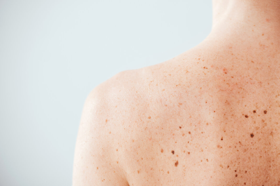
CUTANEOUS AND SUBCUTANEOUS TUMORS

Cutaneous and subcutaneous tumors are medical conditions in which the cells of the skin and subcutaneous tissues grow abnormally. These tumors can be benign or malignant and can appear in different parts of the body.
Skin tumors include several types of tumors, including basal cell carcinomas (Basal cell carcinoma, the most common skin tumor), squamous cell carcinomas (Squamous cell carcinoma, the second most common skin tumor), and melanomas (Malignant Melanoma, the most dangerous carcinoma). Subcutaneous tumors, on the other hand, may include lipomas, fibromas, and neurofibromas.
Cutaneous and subcutaneous tumors are often caused by a combination of environmental and genetic factors, such as exposure to sunlight, age, sex, family history, and immunodeficiency. Some of these tumors can be prevented with proper sun protection and early diagnosis.
The diagnosis of cutaneous and subcutaneous tumors is made through a biopsy, in which a tissue sample is taken for microscopic analysis. Treatment depends on the type, location, and severity of the tumor and may include surgical removal, radiotherapy, or chemotherapy.
THE SURGICAL TREATMENT is one of the most effective ways to remove skin tumors.
During the surgical procedure, the surgeon removes the tumor along with the surrounding healthy tissue to ensure that the entire tumor has been eliminated. The type of surgical procedure depends on the size and location of the tumor. In plastic surgery, patients can benefit from advanced techniques for the removal of skin tumors.
Surgery can be performed using techniques such as standard excision or excision and reconstruction with flaps and/or grafts, in order to restore the functionality and aesthetics of the treated area. The choice of treatment depends on the characteristics of the tumor and its location.
Post-operative recovery is usually brief and patients can return to their daily activities within a few days. However, it is important to follow the surgeon's instructions to ensure proper healing and prevent complications.
It is important to note that many skin and subcutaneous tumors are curable if diagnosed and treated early. For this reason, it is advisable to consult your doctor if you notice suspicious skin lesions or changes in the color, shape, or size of an existing mole.
BASAL CELL CARCINOMA (BASALIOMA)
THE MOST COMMON SKIN CARCINOMA
Basal cell carcinoma (BCC) is the most common form of skin cancer and, among all types of cancer, it is the most frequently found. Over 45,000 new cases are diagnosed each year. BCCs arise following abnormal and uncontrolled growth of basal cells in the epidermis, one of the three main types of cells in the upper layer of the skin.
Since growth is slow, most BCCs are curable and cause only local damage if diagnosed and treated early. Understanding the causes, risk factors, and warning signs of BCC can help with early diagnosis. The earlier the diagnosis, the easier they are to treat and cure.
Clinical presentation of Basal Cell Carcinoma
BCCs present as open sores (ulcus rodens), red spots, pink growths, shiny bumps, scars, or growths with slightly raised and rounded edges and/or a central indentation. Sometimes basal cell carcinomas can become superinfected, form scabs (crusts), itch, or bleed. The lesions generally, but not exclusively, appear in areas of the body exposed to sunlight. In patients with darker skin, about half of basal cell carcinomas are pigmented (that is, brown in color).
It is important to note that basal cell carcinomas can appear quite different from one individual to another.
90% of skin cancers other than melanoma (mainly Basal cell carcinomas, 75%, and Squamous cell carcinomas, 20%) are associated with exposure to UV rays from the sun.
Are Basal Cell Carcinomas dangerous?
Although BCCs rarely spread beyond the site of the primary tumor, if left to grow, they can give rise to disfiguring and dangerous lesions. Untreated basal cell carcinomas can become locally invasive, expanding in width and depth into the skin and destroying skin, tissues, and bones. The longer you wait before starting to treat a basal cell carcinoma, the more likely it is to recur, sometimes multiple times.
Basal cell carcinoma that spreads to other parts of the body becomes particularly aggressive. In even rarer cases, this type of basal cell carcinoma can become potentially fatal.
Early diagnosis
Thanks to early diagnosis and treatment, almost all basal cell carcinomas (BCC) can be successfully removed without complications. Scheduled visits with the Dermatology Specialist and self-examination, checking for any new lesions with an unusual appearance, allow for the detection of skin carcinomas such as Basal Cell Carcinoma when they are easier to treat.
Basal Cell Carcinoma Diagnosis: Five Warning Signs
Check for the presence of basal cell carcinomas where the skin is more exposed to the sun.
They will often look like this:
1. An open ulcer that does not heal and may bleed, become infected, or crusty
The lesion may last for weeks, or seem healed and then return.
2. A reddened or irritated area on the face, chest, shoulders, arms, or legs that may itch, be sore, or cause no discomfort at all.
3. A shiny swelling or a nodule with a pearly or translucent appearance, pink, red, or whitish.
The swelling can also be dark, black or brown, especially in people with dark skin, and can be mistaken for a normal mole.
4. A small pinkish growth with a slightly raised and rounded edge and a scaly indentation in the center that may develop small superficial blood vessels over time.
5. An area similar to a scar that is flat and white, yellow, or pale in color. The skin appears shiny and tight, often with poorly defined edges. It can often indicate an invasive Basal Cell Carcinoma.
Basal cell carcinomas can be difficult to diagnose
Remember that basal cell carcinomas can also appear different from how they are described. In some individuals, basal cell carcinomas may resemble non-cancerous skin conditions such as psoriasis or eczema.
In case of doubt, have a check-up with your trusted Dermatologist.
What can be done
If you have already suffered from basal cell carcinomas, there is a higher probability of developing another one, particularly in the same area damaged by the sun or nearby.
A basal cell carcinoma can recur even if it was carefully removed the first time, because some cancer cells may not be detected after surgery.
For this reason, it is important to have at least one check-up with your trusted dermatologist once a year.
Follow up: if you have already had a basal cell carcinoma (BCC) or squamous cell carcinoma (SCC) or a precancerous actinic keratosis, it is advisable to consult your doctor periodically according to their recommendations.
Pay attention to sun exposure all year round: avoid unprotected exposure to UV rays, stay in the shade, especially when the sun is strongest, and use a broad-spectrum sunscreen, a wide-brimmed hat, and sunglasses with UV protection.
SQUAMOUS CELL CARCINOMA (SPINALIOMA)
THE SECOND MOST COMMON SKIN CARCINOMA
Squamous cell carcinoma (SCC) of the skin, or spinocellular carcinoma, is the second most common form of skin cancer, characterized by abnormal and accelerated growth of squamous cells. If detected early, most squamous cell carcinomas are curable.
Skin spinocellular carcinomas are also known as cutaneous squamous cell carcinomas (SCC).
How does Squamous Cell Carcinoma present itself?
Squamous cell carcinomas can appear as red scaly patches, open ulcers, with a verrucous appearance or raised exophytic bumps with a central depression. In other cases, squamous cell carcinomas can form superficial crusts, itch, or bleed. The lesions most often arise in areas of the body exposed to the sun.
Squamous cell carcinomas can also appear in other areas of the body, including the genitals.
How dangerous is SCC?
While most Squamous Cell Carcinomas can be treated easily and successfully, if not addressed early, these lesions can become disfiguring, dangerous, and even fatal. Untreated squamous cell carcinomas tend to be invasive, both locally and systemically; they can grow into the deeper layers of the skin and spread to other parts of the body.
Diagnosis of Spinalioma: warning signs
In the more advanced stages, these skin tumors can become dangerous.
Squamous cell carcinoma of the skin can develop anywhere on the body, but is most often found in areas exposed to ultraviolet (UV) radiation such as the face, lips, ears, scalp, shoulders, neck, backs of the hands, and forearms. Squamous cell carcinomas can develop on scars, skin sores, and other areas of skin injury (Marjolin's ulcer). The surrounding skin typically shows signs of sun exposure damage such as wrinkles, pigment changes, and loss of elasticity.
Squamous cell carcinomas can appear as thickened, rough, scaly areas that may form crusts or bleed. They can also have a warty appearance or sores that never fully heal. In some cases, SCCs present as exophytic growths with raised edges and a typical ulcer or depression in the center that may bleed or itch.
What you can do
It is absolutely important to have at least one check-up with your trusted dermatologist once a year.
Follow up: if you have already had a basal cell carcinoma (BCC) or squamous cell carcinoma (SCC) or a precancerous actinic keratosis, it is advisable to consult your doctor periodically according to their recommendations.
Pay attention to sun exposure all year round: avoid unprotected exposure to UV rays, stay in the shade, especially when the sun is strongest, and use a broad-spectrum sunscreen, a wide-brimmed hat, and sunglasses with UV protection.
THE MOST DANGEROUS CARCINOMA
Cutaneous melanoma is a tumor that originates from the malignant transformation of melanocytes, some of the cells that make up the skin. Melanocytes, together with keratinocytes, are part of the epidermis and are responsible for producing melanin, a pigment that protects against the harmful effects of UV rays. Under normal conditions, melanocytes can form dark clusters visible on the surface of the skin, known as moles (nevi is the medical term).
Cutaneous melanoma is quite rare in children, it mainly affects people around 45-50 years old, although in recent decades, a trend towards a lower average age at diagnosis has been observed.
In Italy, the incidence in the last 10 years has doubled with 7,300 new cases each year among men and 6,700 among women
Cutaneous melanoma represents only a small percentage (about 5%) of skin cancers.
Risk Factors
The main risk factor for cutaneous melanoma is excessive exposure to ultraviolet light (UVA and UVB rays) (sun rays, lamps, etc.).
Other known risk factors are immune system deficiency (following chemotherapy or transplants) and some hereditary diseases, such as xeroderma pigmentosum, in which DNA is unable to repair damage caused by radiation. The risk also increases in people with freckles or many moles, in those with light eyes, hair, and skin, and in those who have a close relative affected by this tumor or who have had a previous cutaneous melanoma.
Types of Melanoma
Cutaneous melanomas originate either on intact skin or from pre-existing nevi, which are present from birth or early childhood (congenital) or appear during the course of life (acquired).
From a clinical point of view, four types of cutaneous melanoma are distinguished: superficial spreading melanoma (the most common, accounting for about 70 percent of all cutaneous melanomas), lentigo maligna melanoma, acral lentiginous melanoma, and nodular melanoma (the most aggressive, accounting for about 10-15 percent of cutaneous melanomas).
Unlike the first three types, which initially have a superficial growth, nodular melanoma invades the tissue in depth from its earliest stages.
Warning symptoms
The main sign of cutaneous melanoma is a change in the appearance of a pre-existing mole or the appearance of a new one. The characteristics of a mole that may indicate the onset of melanoma are summarized by the acronym ABCDE:
A for Asymmetry in shape (a benign mole is generally circular or rounded, a melanoma is more irregular);
B for Irregular and indistinct Borders;
C for Variable Color (that is, with different shades within the mole itself);
D for Increasing Dimensions, both in width and thickness;
E for Evolution of the mole which, in a rather short time, shows changes in appearance.
Other warning signs that should be evaluated by a doctor are a mole that bleeds, itches, or is surrounded by a nodule or a reddened area.
It is essential to ensure early treatment of the disease, because, if not treated promptly, Cutaneous Melanomas have an invasive course and lead to a poor prognosis.
What you can do
It is absolutely important to have at least one check-up with your trusted dermatologist once a year.
Follow up: if you have already had a Cutaneous Melanoma carcinoma, it is advisable to consult your doctor periodically according to their recommendations.
Pay attention to sun exposure all year round: avoid unprotected exposure to UV rays, stay in the shade, especially when the sun is strongest, and use a broad-spectrum sunscreen, a wide-brimmed hat, and sunglasses with UV protection.
For more information
